Dry Age-Related Macular Degeneration

Approximately 85% to 90% of the cases of macular degeneration are the dry (atrophic) type. Dry age-related macular degeneration does not involve any leakage of blood or serum. Loss of vision may still occur. Patients with this dry form may have good central vision (20/40 or better) but substantial functional limitations, including fluctuating vision, difficulty reading because of their limited area of central vision, limited vision at night or under conditions of reduced illumination.

In the dry type of macular degeneration, the deterioration of the retina is associated with the formation of small yellow deposits, known as drusen, under the macula. This phenomenon leads to a thinning and drying out of the macula, causing the macula to lose its function. The amount of central vision loss is directly related to the location and amount of retinal thinning caused by the drusen.

The early stage of dry age-related macular degeneration is associated with minimal visual impairment and is characterized by large drusen and pigmentary abnormalities in the macula. Drusen are accumulations of acellular, amorphous debris subjacent to the basement membrane of the retinal pigment epithelium. Nearly all people over the age of 50 years have at least one small druse in one or both eyes. Only eyes with large drusen are at risk for late age-related macular degeneration.

This form of macular degeneration is much more common than the “wet” type of macular degeneration, and it tends to progress more slowly than the “wet” type. However, a certain percentage of the “dry” type of macular degeneration turns to “wet” with the passage of time. There is no known cure for the “dry” type of macular degeneration.

Wet Age-Related Macular Degeneration

Approximately 10-15% of the cases of macular degeneration are the “wet” (exudative) type.

In the “wet” type of macular degeneration, abnormal blood vessels (known as choroidal neovascularization or CNV) grow under the retina and macula. These new blood vessels may then bleed and leak fluid, causing the macula to bulge or lift up from its normally flat position, thus distorting or destroying central vision. Under these circumstances, vision loss may be rapid and severe.

With the “wet” type, patients may see a dark spot (or spots) in the center of their vision due to blood or fluid under the macula. Straight lines may look wavy because the macula is no longer smooth. Side or “peripheral” vision is rarely affected. However, some patients do not notice any such changes, despite the onset of neovascularization . Therefore, periodic eye examinations are still very important for patients at high risk.

There are two forms of choroidal neovascularization (CNV) that have been identified, “classic” and “occult.” The classic form is well-defined and usually results in vision that is between 20/250 and 20/400, but it may be worse than 20/800. For eyes with the occult form, the average visual acuity is somewhat better, between 20/80 and 20/200. Occult lesions are not well-delineated and they have less leakage.

Once CNV has developed in one eye, whether there is a visual loss or not, the other eye is at relatively high risk for the same change. When all four risk factors—more than five drusen, large drusen, pigmental clumping, and systemic hypertension—are present, the five-year risk of CNV in the second eye is 87%, whereas if none of these risk factors are present, the risk is 7%.

In addition, CNV may progress rapidly, and any sudden change in central vision therefore requires a prompt examination after dilation of the eyes. The purpose of this exam is to find out whether the sudden loss of vision is due to leakage of blood vessels and which treatment may be appropriate.

Reducing your Intraocular Pressure may not be enough to stop your Glaucoma progression!

Today, IOP management remains the standard of care, even though IOP elevation is not pathognomonic of glaucoma, and patients can continue to lose vision despite effective IOP control.

Our breakthrough and revolutionized Netra Restoration Therapy (NRT) will provide Neuroprotection, Reduce Ocular Inflammation, Oxidative Stress, Restore Normal Blood flow and supply Antioxidants to your eyes to protect and restore your vision.

Find out if you are a candidate for NRT.

What is Keratoconus?

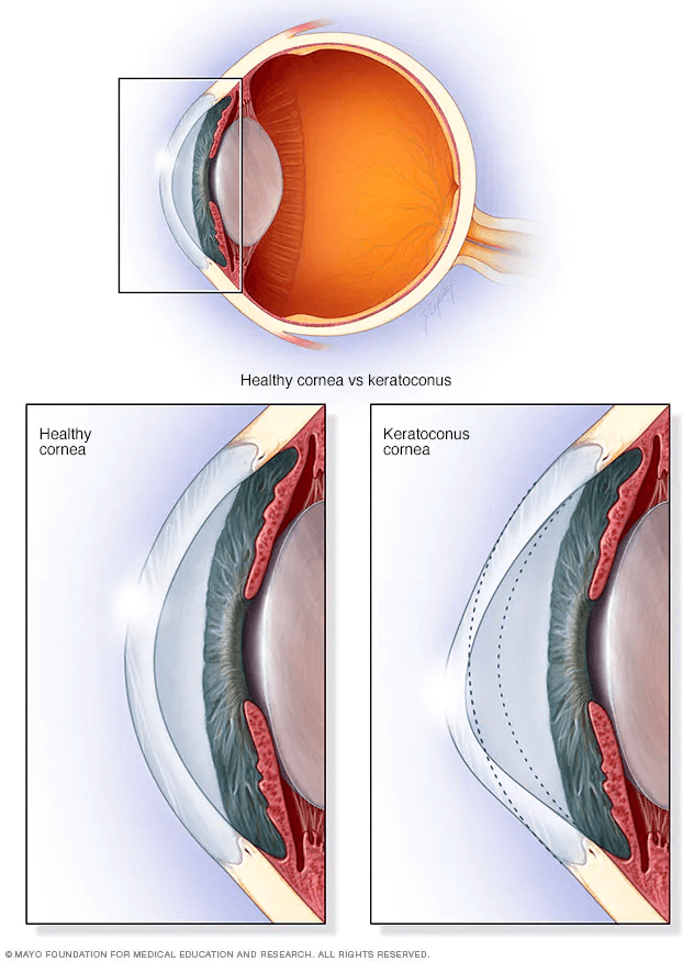
Keratoconus is an eye condition that causes the cornea to become progressively thinner. A normal cornea is round or spherical in shape, but with keratoconus the cornea bulges forward, assuming more of a cone shape. As light enters the cone shaped cornea it is bent and distorted and unable to come to a point of clear focus on the light-sensitive retina.

Keratoconus usually affects both eyes but the two eyes often progress at different rates. This disease typically begins during teenage years. In most patients, it progresses for several years before stabilizing in the third to fourth decade of life. In severe cases it can continue to worsen. In these cases the cornea continues to thin and bulge outward, further blurring vision. Scarring of the cornea can also develop.

When people with diabetes experience long periods of high blood sugar, fluid can accumulate in the lens inside the eye that controls focusing. This changes the curvature of the lens, leading to changes in vision. However, once blood sugar levels are controlled, usually the lens will return to its original shape and vision improves. Patients with diabetes who can better control their blood sugar levels will slow the onset and progression of diabetic retinopathy.

How do we treat Keratoconus at Netra?

Acupuncture

Clinical studies have shown Acupuncture is effective in a wide range of disorders including Eye Diseases, Diabetes, Hyperthyroidism, Hypothyroidism, Hashimoto, Cushing’s Syndrome, Osteoporosis, Thyroiditis, PCOS / Addison’s Disease and Menopause.

Increases Ocular Circulation.

Reduces Inflammation and Increases Circulation.

Non-Surgical and Opioid-Free treatment.

No side effects and safe.

Releases Endorphins to combat pain.

Ayurvedic Medicine

Ayurveda offers one of the most effective medicines for digestive issues and helps to correct root cause of your condition.

Complete holistic healing system in existence more than 3000 years.

Based on the concept of root-cause diagnosis and management.

Focuses equally on prevention and cure to improve quality of life.

Helps identify and recommends foods for your body type and condition.

Herbal Medicine

Herbal medicine has a history of at least several thousand years and uses mostly plants to treat diseases and promote health.

Can successfully treat many chronic and complex conditions.

Clinically and scientifically proven to treat a wide range of complex eye conditions.

Safe and has relatively less side effects.

Helps boost your immune system naturally.

Shown to reduce stress and relieve anxiety.

Improves respiratory and cardiovascular function.

Therapeutic Yoga

Therapeutic Yoga or Yoga Therapy involves employing a variety of yoga practices to help improve a health.It also adapts the practice of Yoga to the needs of people with specific health condition.

Improves strength, balance and flexibility.

Helps with chronic pain relief.

Improves circulation and reduces blood pressure.

Reduces stress and improves sleep.

Improves respiratory and cardiovascular function.

Symptoms Of Keratoconus:
Who Is At Risk:

REASONS FOR YOUR 
GLAUCOMA PROGRESSION‍

The below factors can also contribute to your Glaucoma progression.

Neuroprotection
Vascular Dysregulation
Oxidative Stress
Ocular Inflammation
Excitotoxicity

TREATMENT BENEFITS

Netra Restoration Therapy provides the following benefits for patients with Glaucoma.
The level of loss of nerve fiber layer and optic nerve atrophy will determine the level of the improvement seen.

Results Visible in 7 Days
Stops Progression
Improved Visual Field
Improved Visual Acuity
Improved Color Contrast
Reduced Bluriness/Glare

RESEARCH ARTICLES SHOWING INSUFFICIENT OCULAR BLOOD FLOW IN GLAUCOMA

Oxygen and blood flow: Players in the pathogenesis of glaucoma

Current evidence supporting the role of ocular blood flow (OBF) and oxidative stress in the pathogenesis of glaucomatous damage.

Clinical clues of vascular dysregulation and its association with glaucoma

Evidence linking vascular dysregulation and glaucoma, and evaluate the clinical characteristics that might suggest the presence of vascular dysregulation in the glaucoma patient.

Blood flow in Glaucoma

Several studies indicate that a perfusion instability, rather than a steady reduction of ocular blood flow, might contribute to glaucomatous optic neuropathy.

The doctor combines the best of Chinese and Indian medicine to heal issues for which there appear to be no other solutions. In my case, I presented with glaucoma with 20/30 vision and a small loss in peripheral vision. The doctor restored my vision to 20/15 and improved the visual field in my left eye so that it is normal and the visual field in my right eye so that it is almost normal.

– Martin

Hear from our patients

INCREASED INTRAOCULAR PRESSURE IS NOT THE ONLY RISK FACTOR FOR GLAUCOMA. ​

Lowering the intraocular pressure with eye drops had been the mainstay for the treatment of Glaucoma.

Increased intraocular pressure is only one of the reason why vision loss happens in glaucomatous eyes.

It is well established that many patients continues to experience Glaucoma disease progression and vision loss despite maintaining lower intraocular pressure.

VISION LOSS EVEN WITH NORMAL IOP

Do you know people with normal intraocular pressure develop Glaucoma and also people with higher-than-normal intraocular pressure never develop Glaucoma.

If you have been treated for Glaucoma through management of intraocular pressure, then you may be addressing only one of the major risk factors and may continue to experience vision loss.

QUESTIONS ?

HOW LONG DO I HAVE STAY  IN CLINIC WHEN I COME FOR TREATMENT?